The human lung performs an important position in respiration, permitting us to breathe in oxygen-rich air whereas expelling carbon dioxide from our our bodies. On this context, it turns into essential to grasp how infections can have an effect on these very important organs.
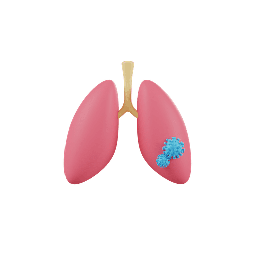
A current examine has unveiled a three-dimensional (3D) illustration depicting the results of the novel coronavirus on the lungs. This progressive visible illustration presents medical professionals and researchers invaluable insights into the development of COVID-19 inside the respiratory system. By inspecting the detailed picture, specialists can higher comprehend the virus’s habits and determine potential therapeutic targets to fight its influence.
The illustration highlights how SARS-CoV-2, the virus chargeable for COVID-19, invades human lung cells by binding to particular receptors on their floor. This interplay triggers an immune response that may result in irritation and harm within the lungs. Because the an infection progresses, it causes the formation of small blood clots inside the pulmonary vasculature, additional impairing oxygen change between the bloodstream and alveoli – tiny sacs chargeable for fuel change within the lungs.
Furthermore, the 3D depiction additionally showcases the influence of extreme instances of COVID-19, the place viral replication happens at such excessive ranges that it overwhelms the physique’s immune defenses. Because of this, sufferers expertise acute respiratory misery syndrome (ARDS), which can require mechanical air flow or different life-saving interventions.
In abstract, the groundbreaking 3D illustration of contaminated lungs offers invaluable info for scientists finding out SARS-CoV-2 and its impact on the respiratory system. With this enhanced understanding, they hope to develop more practical therapies and preventive measures in opposition to future outbreaks of COVID-19 and comparable infectious ailments.


